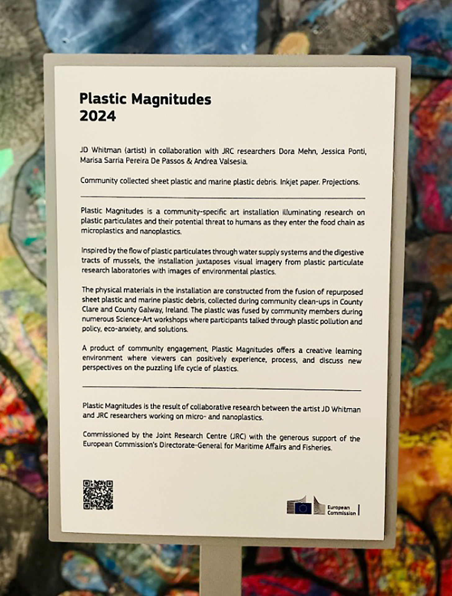
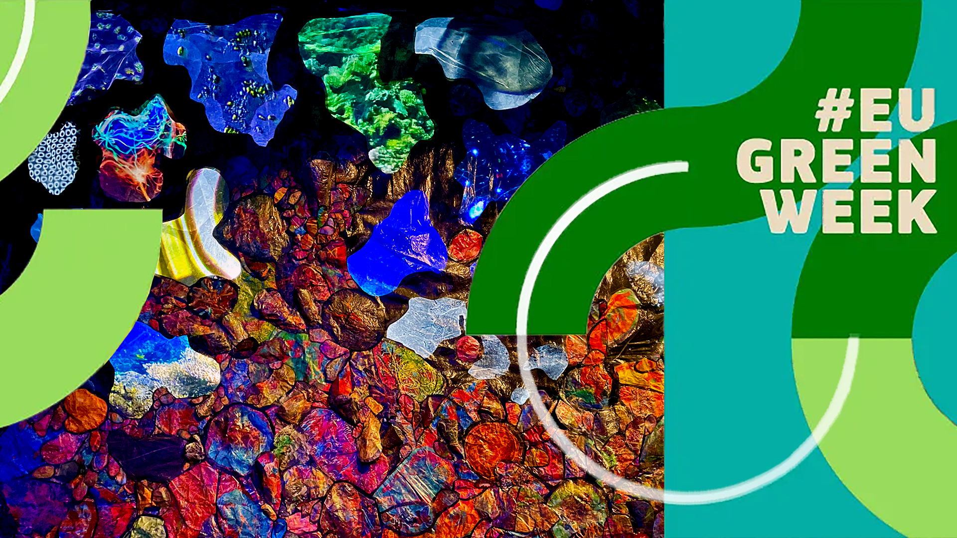
• About
  • PLASTIC MAGNITUDES
  • Plasticity Project
  • BLASCHKA
  • PURGE
  • Portfolio
  • HOME
Menu

JD Whitman

Installation Artist & Ocean Advocate
  • About
  • PLASTIC MAGNITUDES
  • Plasticity Project
  • BLASCHKA
  • PURGE
  • Portfolio
  • HOME

Part of NaturArchy at iMAL - Center for Digital Cultures and Technology. 25.05-29.09.2024

Plastic Magnitudes

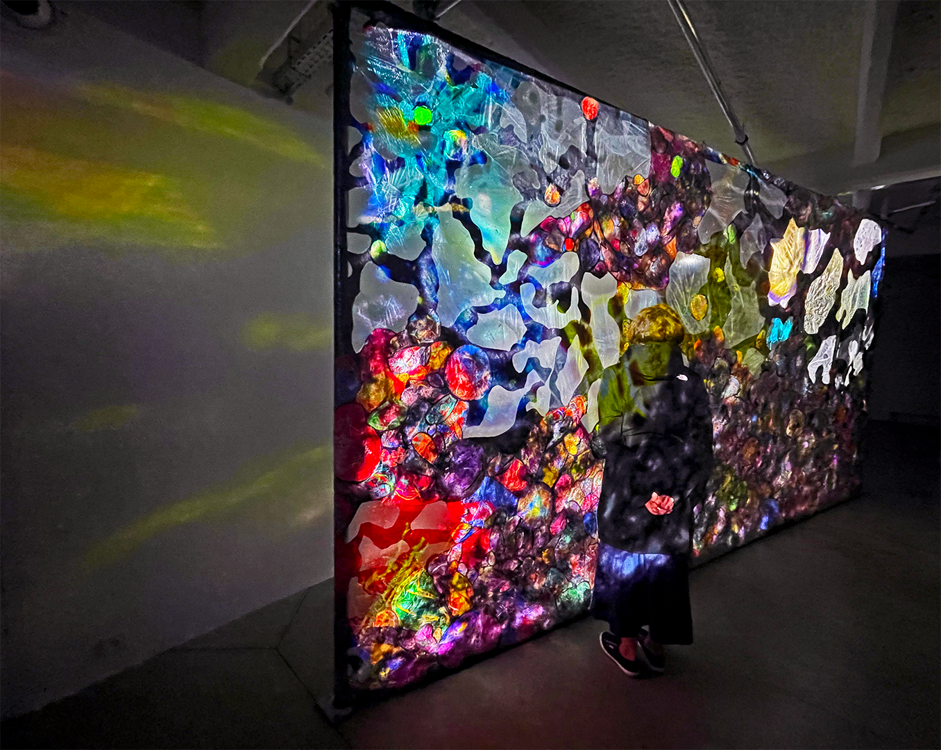
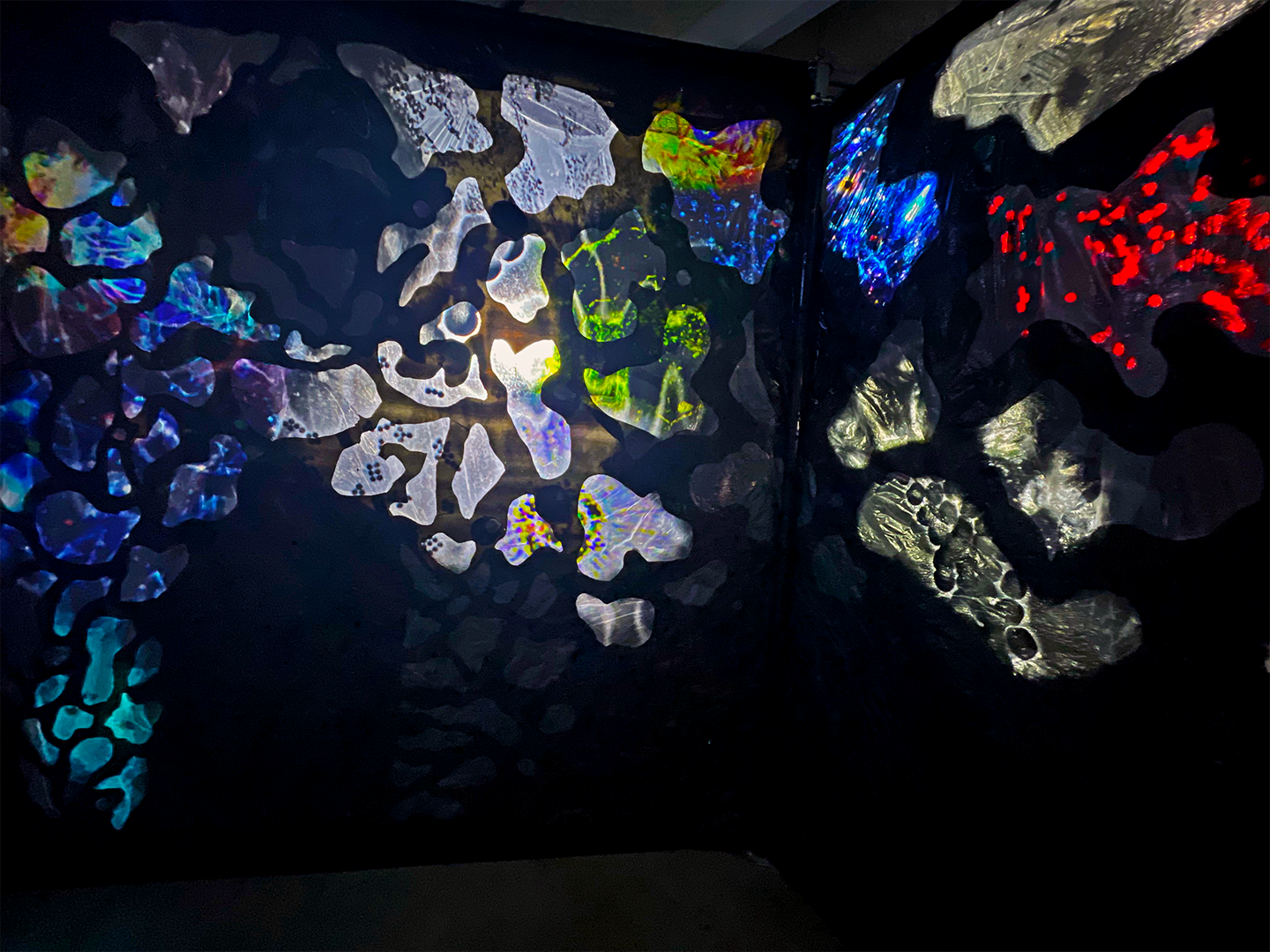
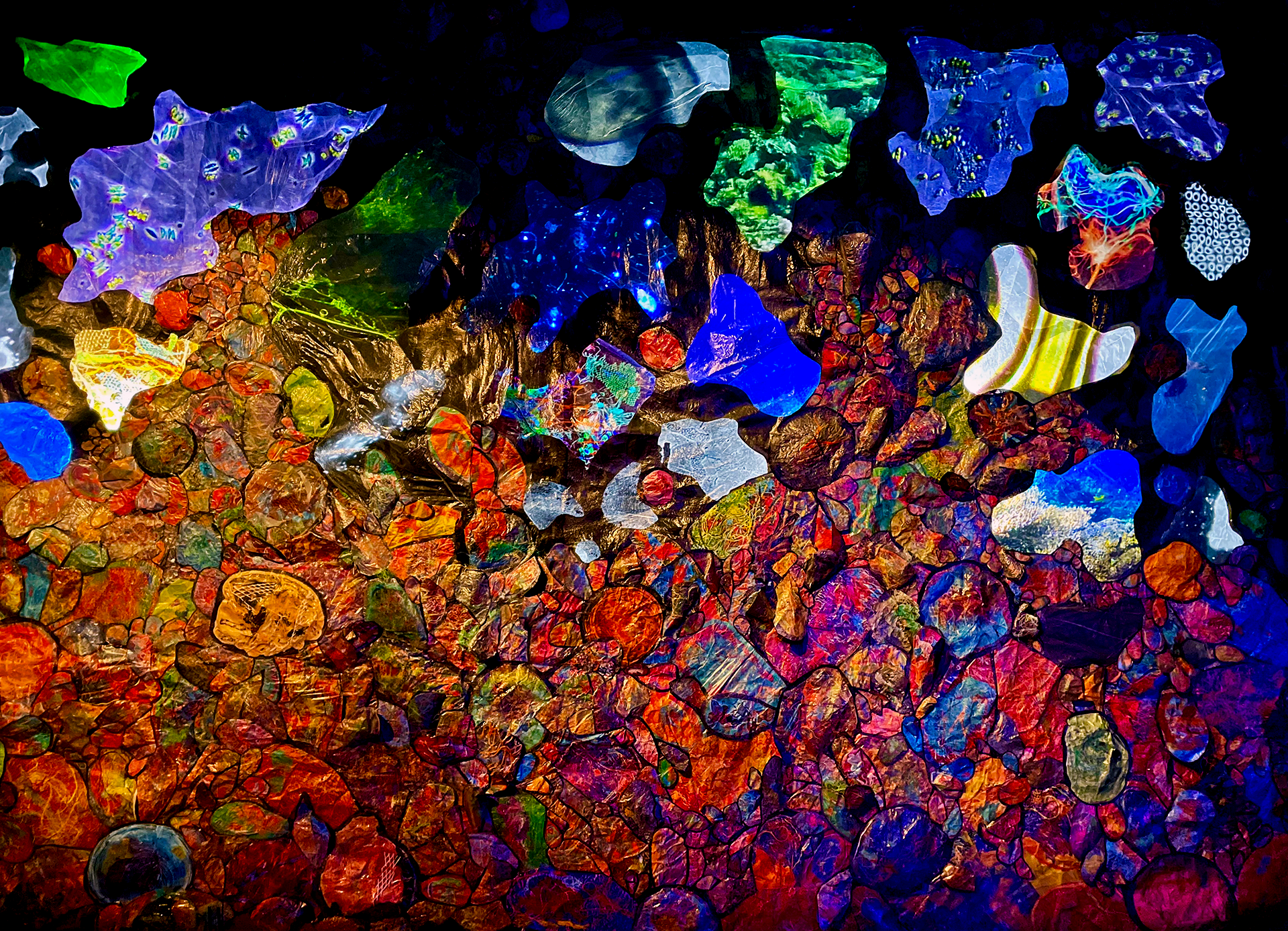
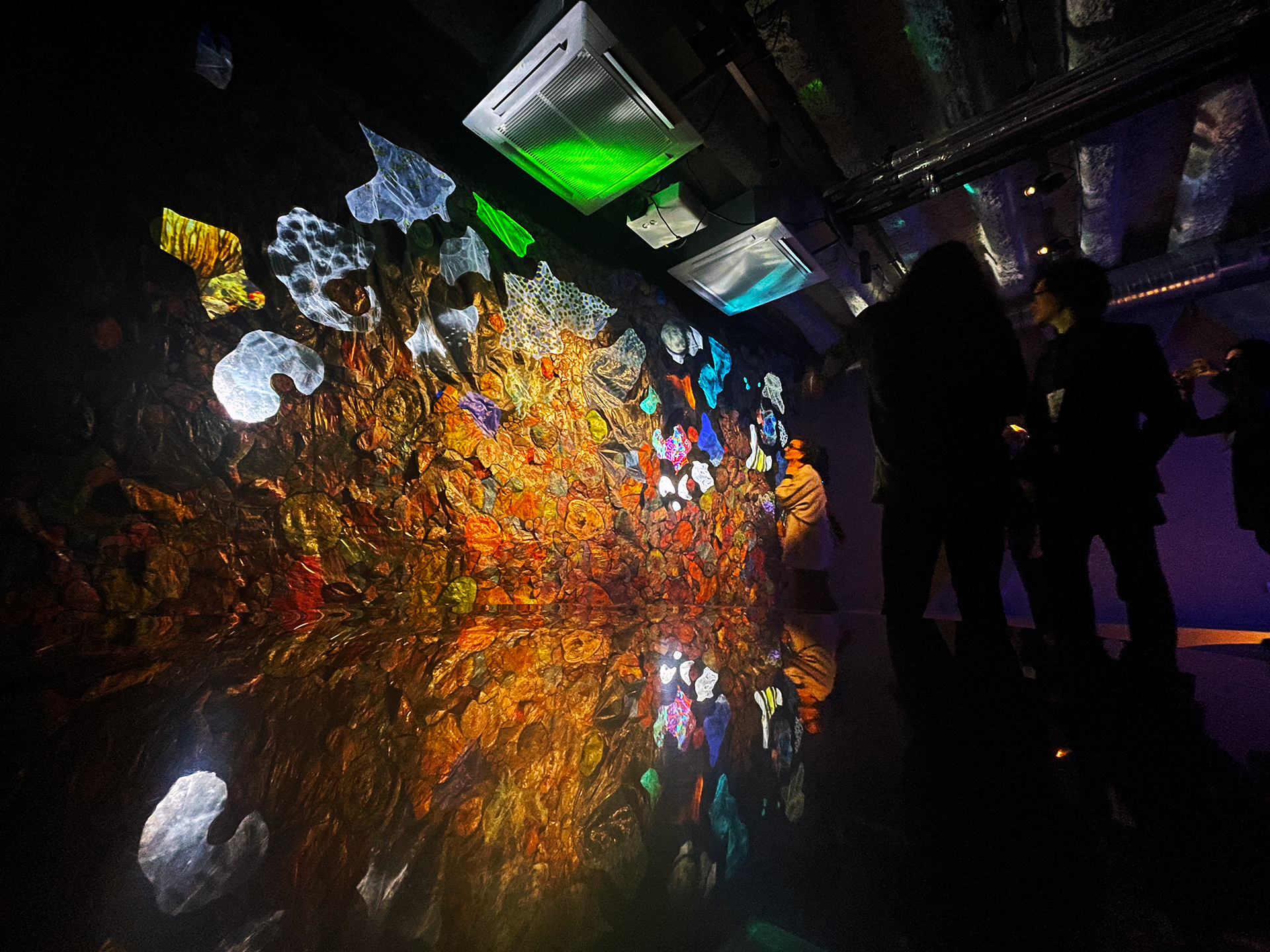
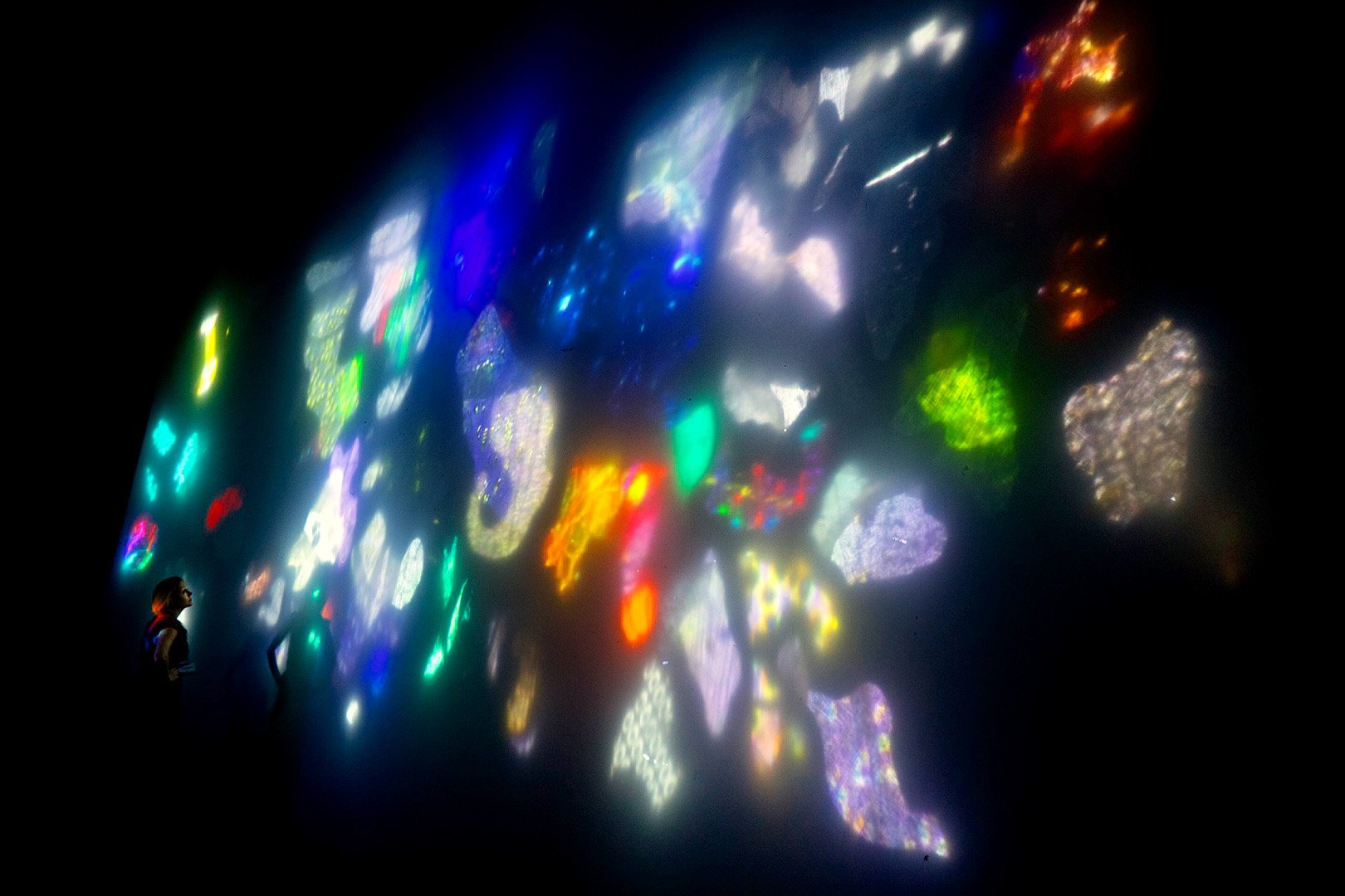
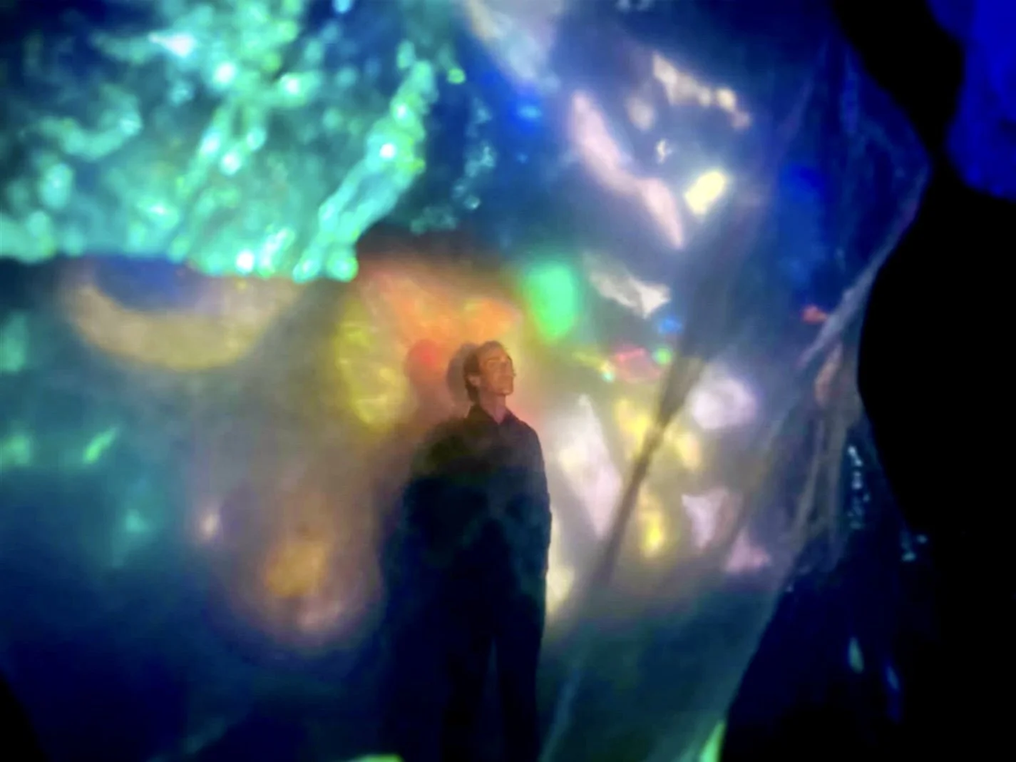
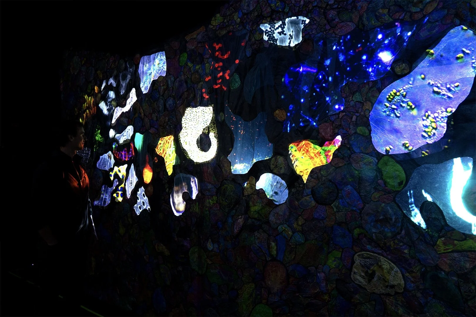
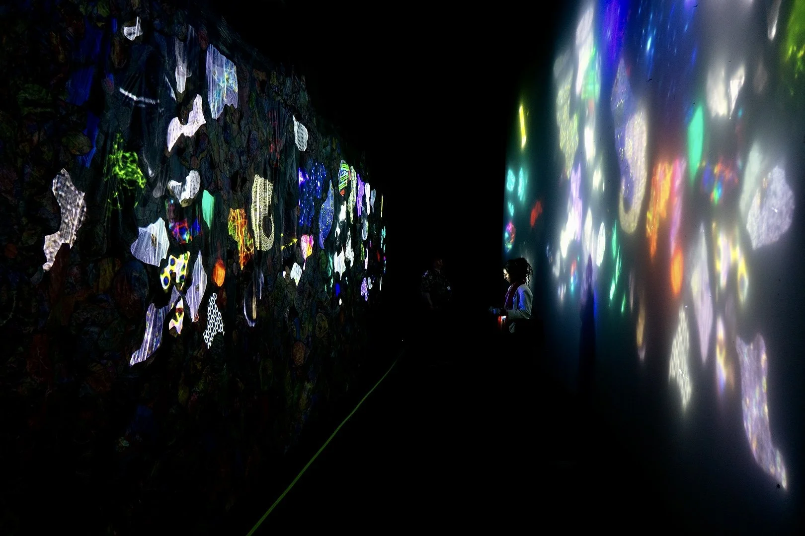
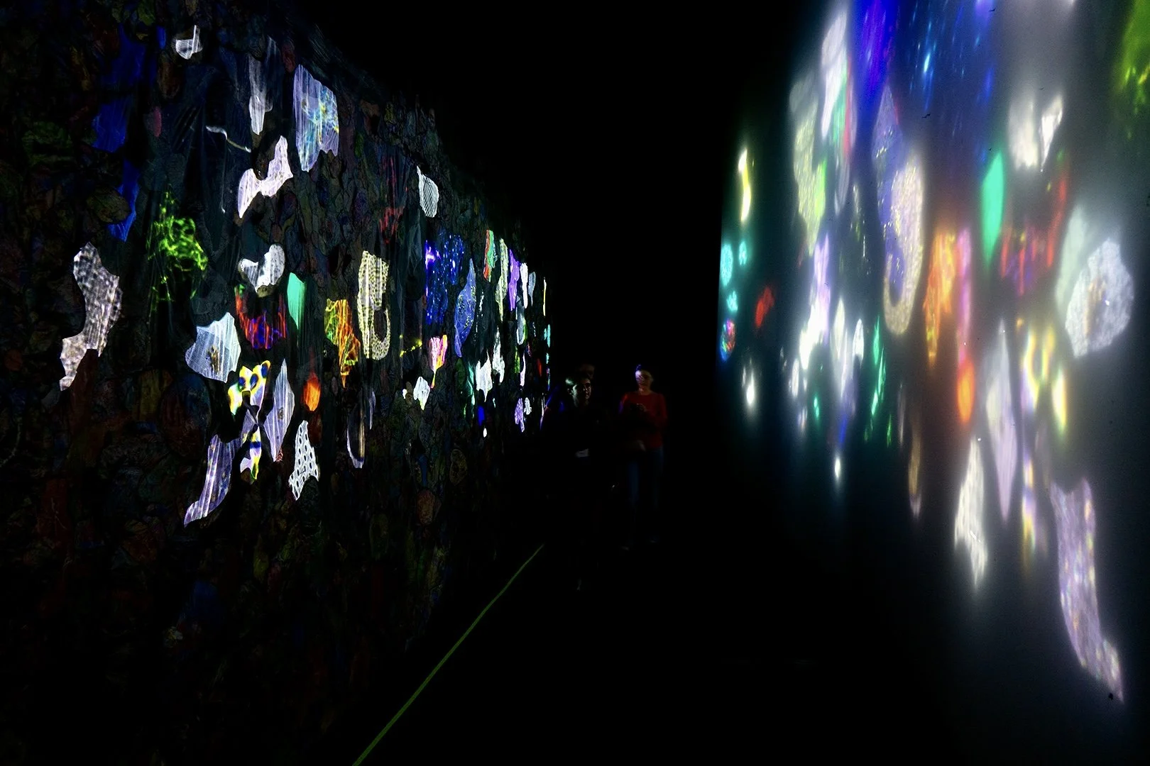
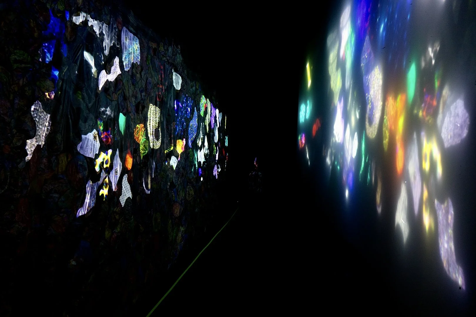
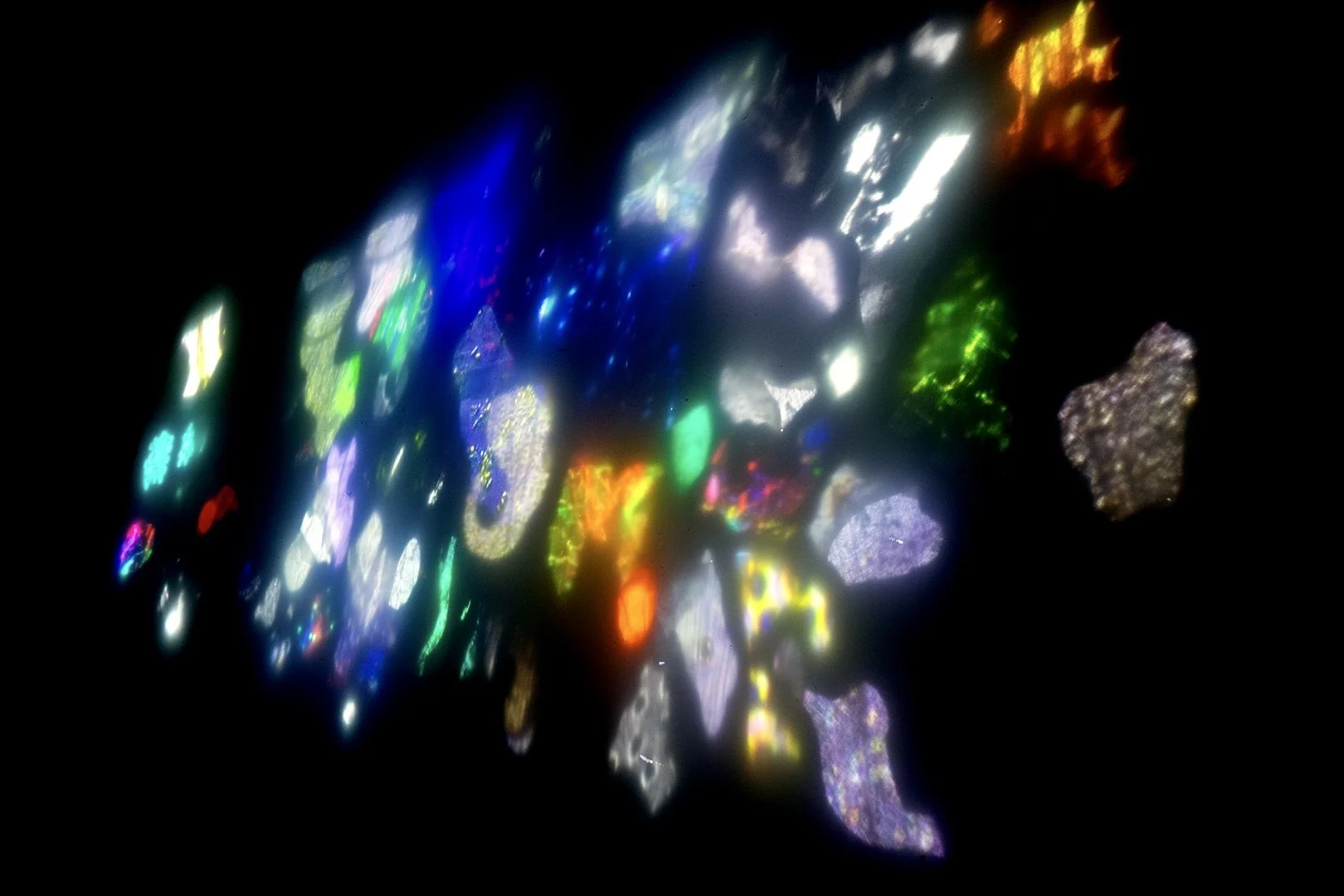
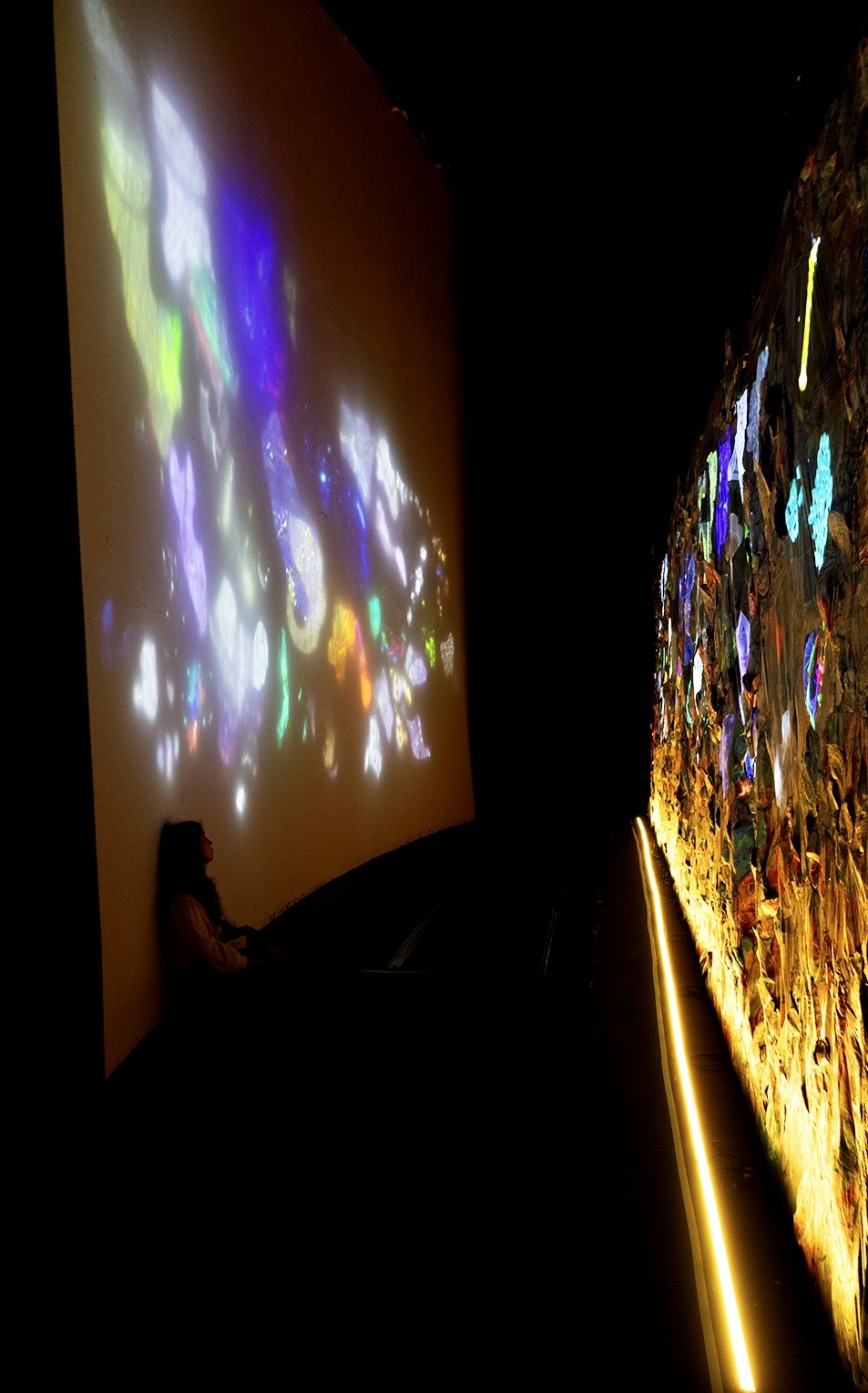
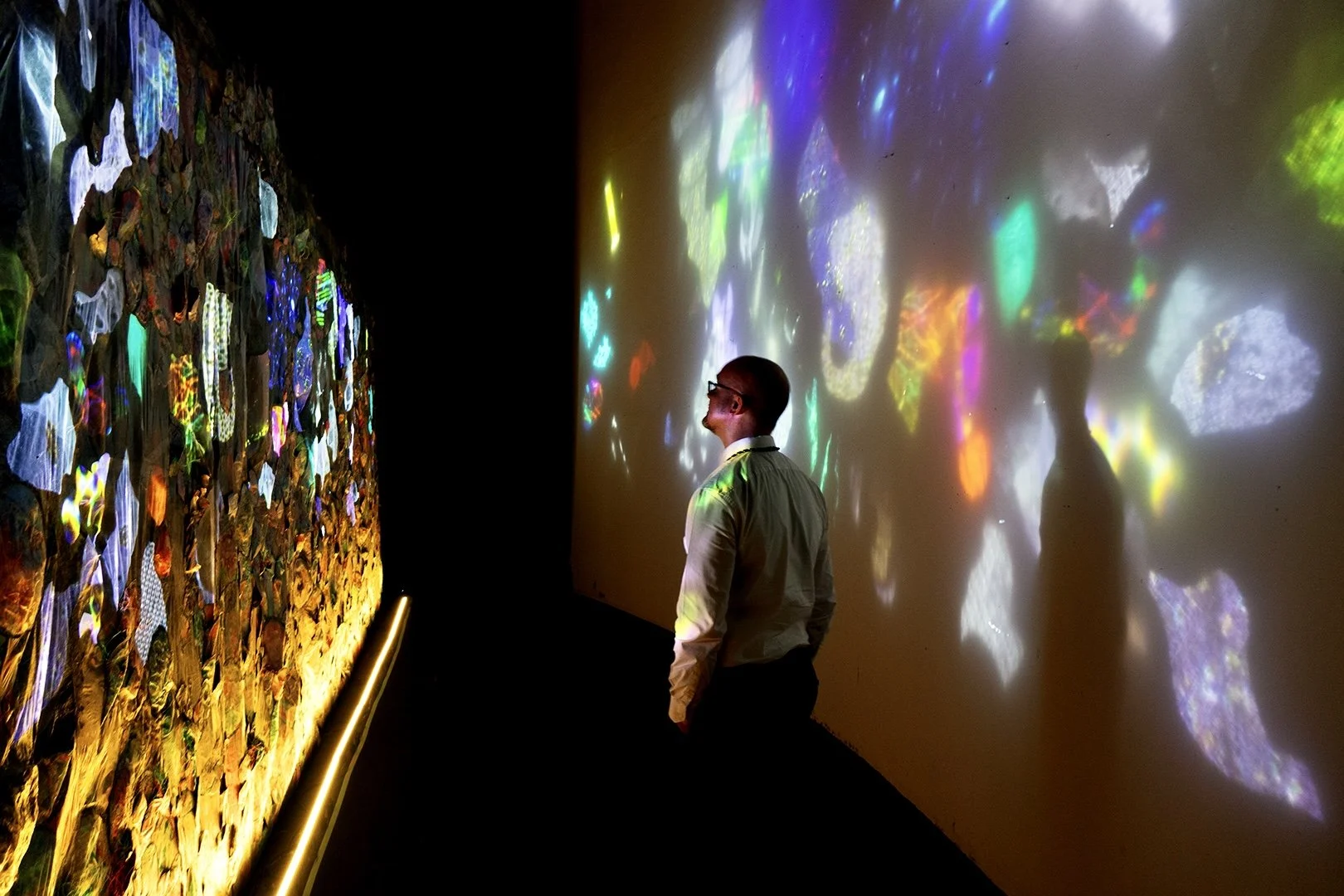
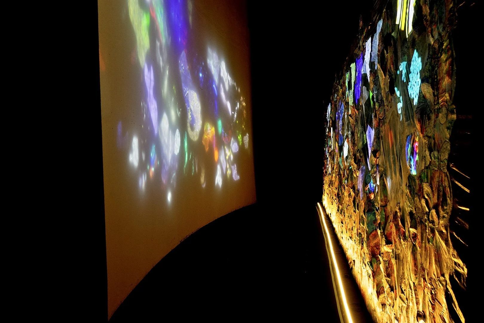
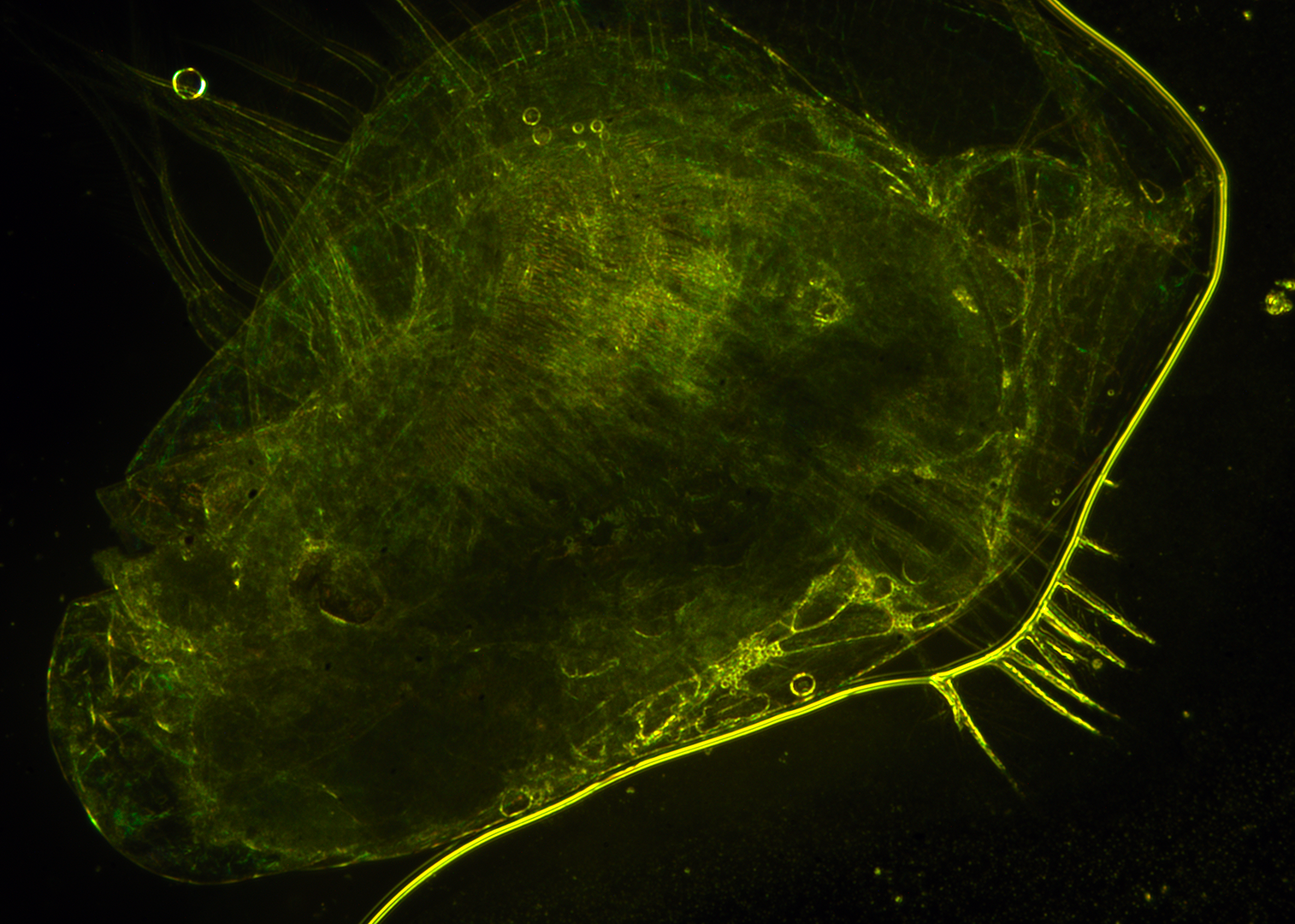
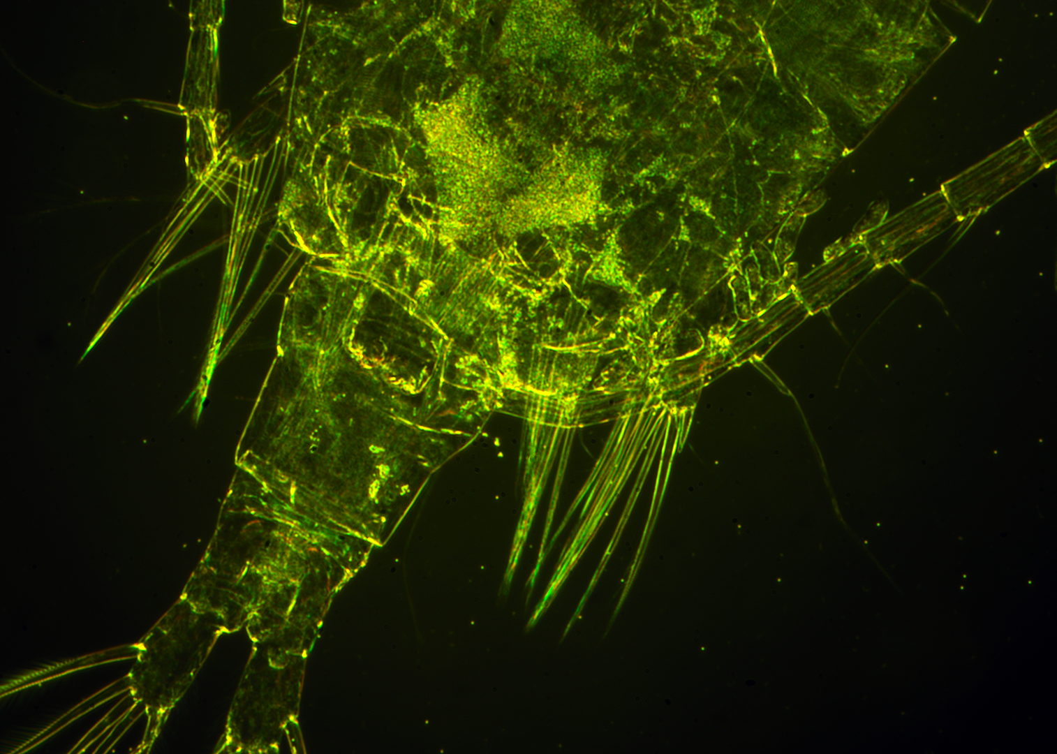
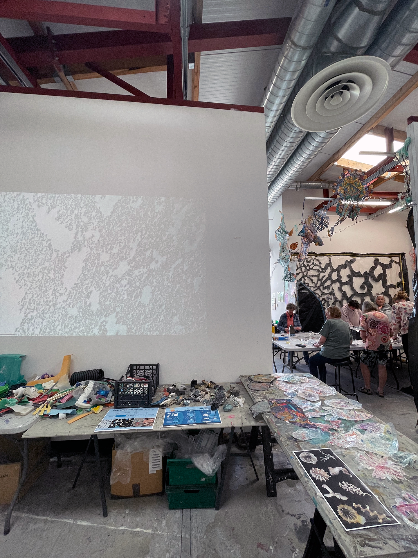
A community-specific, immersive art installation illuminating research from the Joint Research Centre of the European Commission on plastic particulates and their potential threat to humans as they enter the food chain as microplastics and nanoplastics.

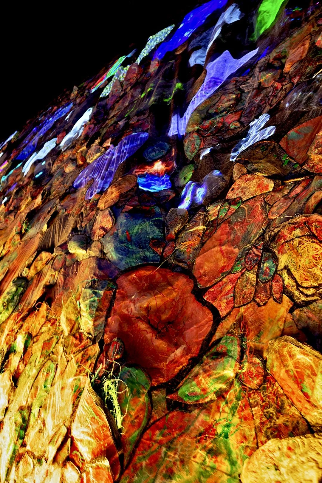
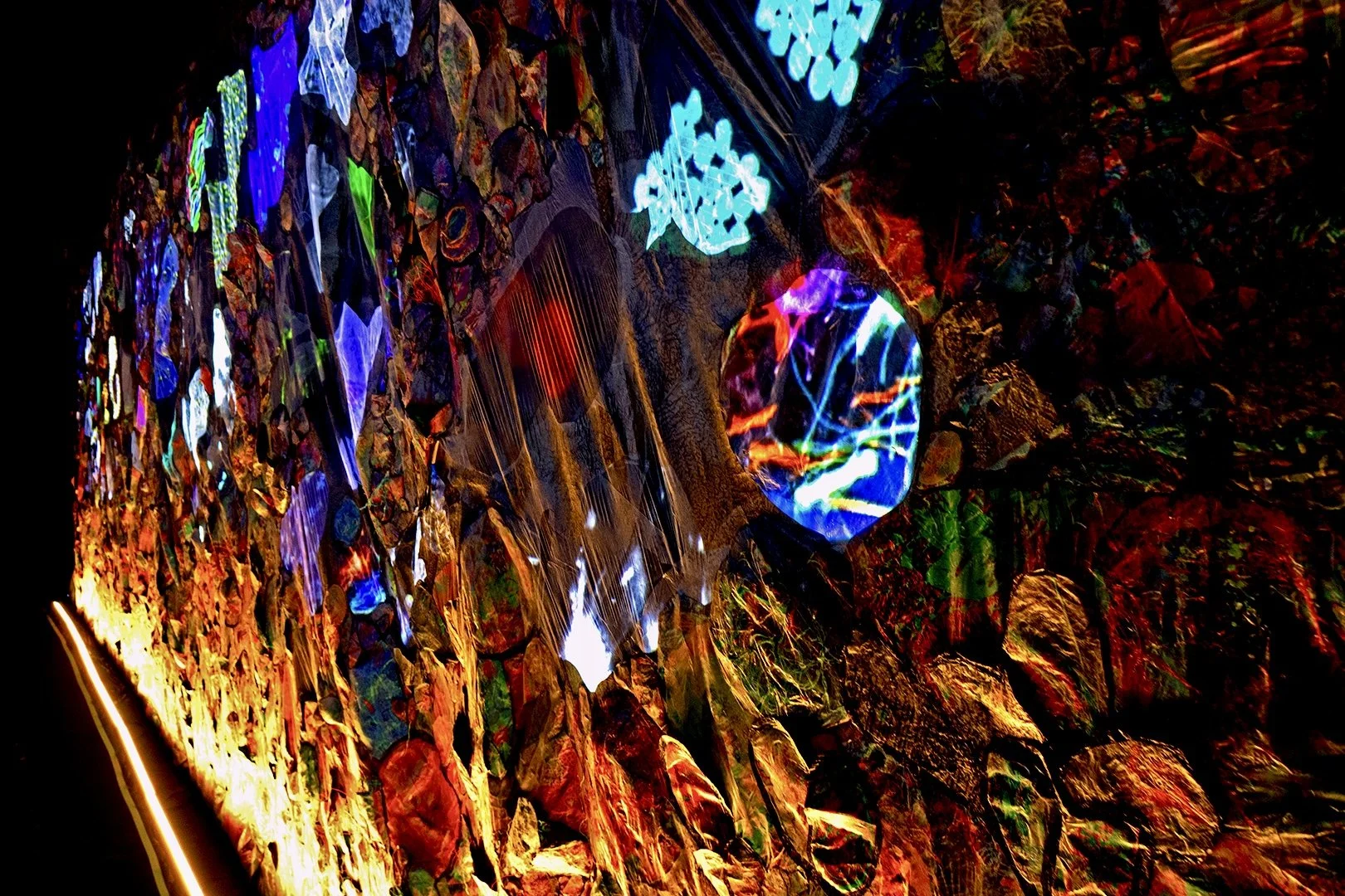
Inspired by the flow of plastic particulates through water supply systems and the digestive tracts of mussels, the installation juxtaposes visual imagery from plastic particulate research laboratories with images of environmental plastics. 

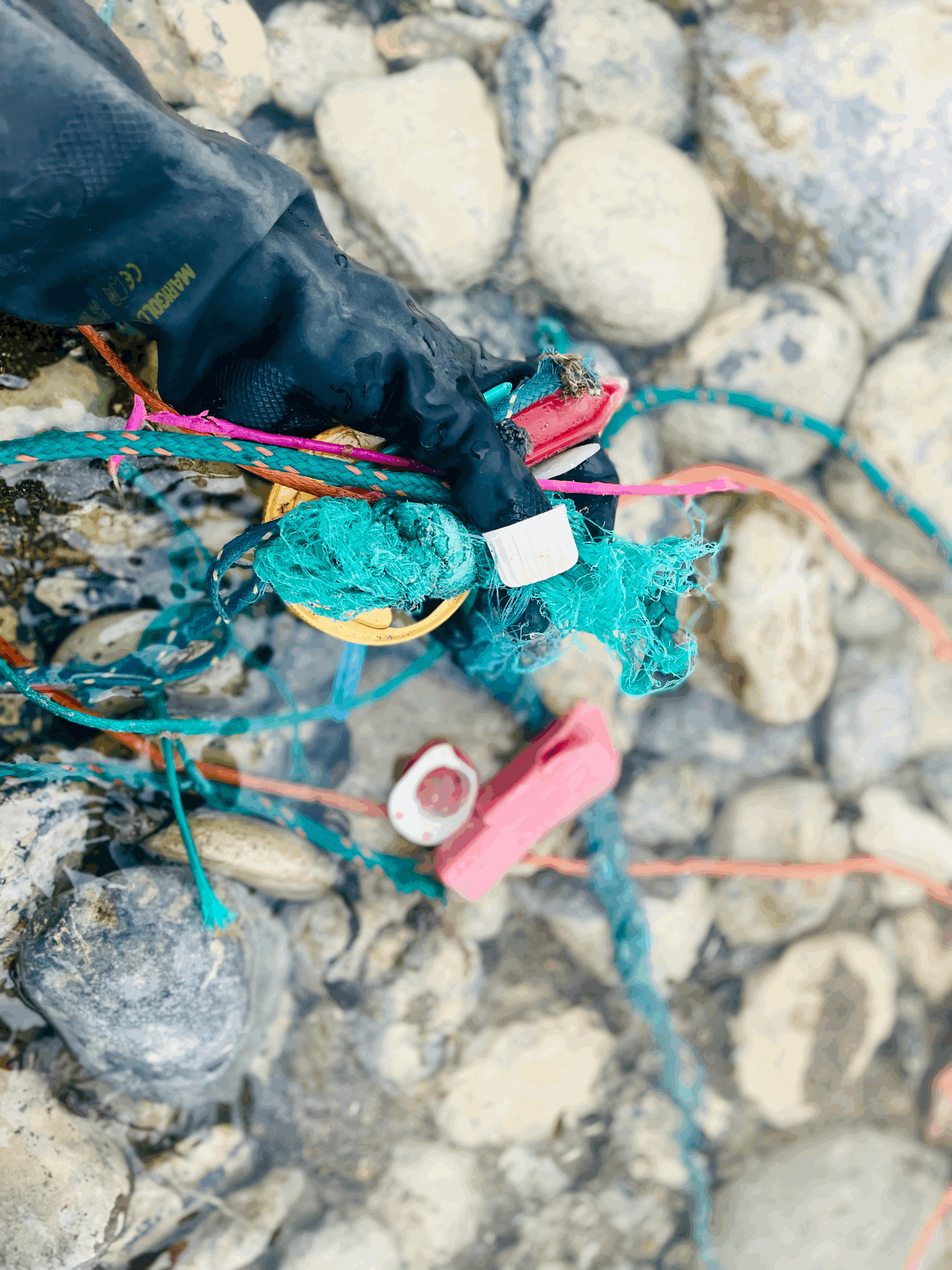
The physical materials in the installation are constructed from the fusion of repurposed sheet plastic and marine plastic debris, collected during community clean-ups in Clare and Galway, Ireland. The plastic was fused together with community members during numerous SciArt workshops where participants talked through plastic pollution and policy, eco-anxiety, and solutions. A product of community engagement, the artwork offers a creative learning environment where viewers can positively experience, process, and discuss new perspectives on the puzzling life cycle of plastics.

ABOUT THE ARTWORK

Plastic Magnitudes

2024

Installation

3.42m x 8.62m x 2.75m

Recycled plastic sheeting, marine plastic debris, inkjet-printed paper, and projected visual imagery derived from JRC research on microplastics and nanoplastics. To learn more about the illuminated JRC research, click HERE.

Collaborating JRC Scientists: Dora Mehn, Jessica Ponti, Marisa Sarria Pereira De Passos & Andrea Valsesia

Program Engineer: Ted Charles Brown

EU Policy Consultant: Danijela Brkovic

Plastic Policy Researcher: Elsa M. K. Brenner

Artist & Workshop Assistant: Lindsay LeBlanc

Developed from 2022-2024 during the fourth edition of Resonances, the flagship initiative of the JRC SciArt Project, following the theme and curatorial concept of NaturArchy: Towards a Natural Contract.

Commissioned for Resonances culminating exhibition - NaturArchy - at iMAL - Center for Digital Cultures and Technology. 25.05 - 29.09.2024


IMG_8592 copy.png
IMG_8669 copy.png
IMG_8583 copy.png
IMG_8523 copy.png
IMG_8679 copy.png
IMG_8575 copy.png
IMG_8553 copy.png
IMG_8559 copy.png
IMG_4529 copy.png
IMG_8565 copy.png
IMG_8596 copy.png
IMG_8561 copy.png
IMG_8595 copy.png
IMG_8709 copy.png
IMG_8719 copy.png
IMG_8723 copy.png
IMG_8591 copy.png
IMG_8623 copy.png
IMG_8610 copy.png
IMG_8629 copy.png
IMG_8675 copy.png
IMG_8502 copy.png
IMG_8603 copy.png
IMG_8611 2 copy.png

ILLUMINATED JRC RESEARCH

Plastic Magnitudes highlights digital materials derived from JRC research on microplastics and nanoplastics.

These materials include images and videos of research methodologies, equipment, and results captured by the project’s collaborating scientists: Dora Mehn, Jessica Ponti, Marisa Sarria Pereira De Passos & Andrea Valsesia.

Each image and video is presented here with a detailed description.

© European Commission

Desmodesmus is a genus of green algae in the family Scenedesmaceae. It is the only chlorophyll-containing organism known to have caused human infections in immunocompetent individuals. All known cases involved open injuries occurring in freshwater. To learn more, read the article: Infection with a Chlorophyllic Eukaryote after a Traumatic Freshwater Injury | New England Journal of Medicine (nejm.org)

© European Commission

A protozoan (most likely an amoeboid) feeding on cells of the green alga Desmodesmus subspicatus.

© European Commission

A protozoan (most likely Favella sp.) feeding on cells of the green alga Desmodesmus subspicatus.

© European Commission

A protozoan (most likely Favella sp.) feeding on cells of the green alga Desmodesmus subspicatus.

© European Commission

Cells of the green alga Desmodesmus subspicatus.

© European Commission

A protozoan (most likely Favella sp.) feeding on cells of the green alga Desmodesmus subspicatus.

© European Commission

Cells of the green alga Desmodesmus subspicatus.

© European Commission

Cells of the green alga Desmodesmus subspicatus.

© European Commission

Microscopic imaging of a water fleas (Daphnia sp.), a small freshwater crustacean that is part of the zooplankton.

© European Commission

Microscopic imaging of a water fleas (Daphnia sp.), a small freshwater crustacean that is part of the zooplankton.

© European Commission

Microscopic imaging of a water fleas (Daphnia sp.), a small freshwater crustacean that is part of the zooplankton.

© European Commission

Microscopic imaging of a water fleas (Daphnia sp.), a small freshwater crustacean that is part of the zooplankton.

© European Commission

Microscopic imaging of a water fleas (Daphnia sp.), a small freshwater crustacean that is part of the zooplankton.

© European Commission

Freshwater algae and Polystyrene particles floating around them.

© European Commission

Heliozoa (commonly known as sun-animalcules) microbial eukaryote (protist) with stiff arms (axopodia) radiating from the spherical body.

© European Commission

Microscopic imaging of Chlorella vulgaris, a green alga highly recognized by their natural chelating competence that makes it among the most used microorganisms in bioremediation studies for mitigation of environmental pollution.

© European Commission

Microscopic imaging of Chlorella vulgaris, a green alga highly recognized by their natural chelating competence that makes it among the most used microorganisms in bioremediation studies for mitigation of environmental pollution.

© European Commission

Cells of the green alga Desmodesmus subspicatus.

© European Commission

Cells of the green alga Desmodesmus subspicatus.

© European Commission

Commercially available, 140 micrometer sized fluorescent polystyrene beads imaged by a fluorescent microscope. These beads are used in recovery studies to assess how many of intentionally added plastic particles can be recovered in a drinking water sampling device.

© European Commission

Particles captured from drinking water using a 20 micrometer cutoff filter, resuspended and collected again on a flat, 0.2 micrometer cutoff aluminum-oxide filter. Visulalised by an optical microscope. Drinking water contains large amounts of naturally occuring particles, including iron oxide, calcium carbonate and silicon oxide.

© European Commission

Particles captured from drinking water using a 20 micrometer cutoff filter, resuspended and collected again on a flat, 0.2 micrometer cutoff aluminum-oxide filter. Visulalised by an optical microscope.

The short, fibre like particles in this case were shown to be silicon oxide, originated from freshwater diatoms.


© European Commission

Particles captured from drinking water using a 20 micrometer cutoff filter, resuspended and collected again on a flat, 0.2 micrometer cutoff aluminum-oxide filter. Visulalised by a fluorescent microscope using UV illumination. Some of the captured particles, including those of natural origin, might be autofluorescent under the UV light. Other fluorescent, fiber like particles might also be present because of background contamination. contamination  often occurs during sampling and sample treatment.

© European Commission

Commercially available, 140 micrometer-sized fluorescent polystyrene beads. Imaged by a fluorescent microscope. These beads are used in recovery studies to assess how many of intentionally added plastic particles can be recovered in a drinking water sampling device.

© European Commission

Commercially available, 140 micrometer sized fluorescent polystyrene beads. Image captured using dark field illumination (optical microscopy). These beads are used in recovery studies to assess how many of intentionally added plastic particles can be recovered in a drinking water sampling device.

© European Commission

Microplastic particles generated from fluorescent highlighter caps under the fluorescent microscope. In-house made, fluorescent model particles might help to test irregular shaped particles in recovery studies, i.e. to assess how many of intentionally added plastic particles can be recovered in a drinking water sampling device or during sample treatment steps.

© European Commission

In-house stained, fluorescent microplastic beads made of polymethyl methacrylate visualized by a fluorescent microscope. Nile red, the dye used here is one of the most often applied molecules to label microplastic particles.

© European Commission

In-house stained, 40 micron size  fluorescent microplastic beads made of polymethyl methacrylate visualized by a fluorescent microscope.

© European Commission

Autofluorescent zooplankton under the fluorescent microscope in UV illumination. These little arthropodes were captured by the filtration system of a drinking water production plant that produces drinking water from lake water.

© European Commission

Transmission Electron Microscopy image of an ultra-fine slice of freshwater algae exposed to nanoparticles. Nanoplastics are not internalized by algae, but can slightly interact with cell walls.


© European Commission

Transmission Electron Microscopy image of an ultra-fine slice of freshwater algae exposed to nanoparticles. Nanoplastics are not internalized by algae, but can slightly interact with cell walls.


© European Commission

Transmission Electron Microscopy image of ultra-fine slice of murine macrophage exposed to in-house synthesized gold labeled Polyvinyl Chloride nanoparticles. Nanoplastics are internalized and stored inside endosomes. To learn more, read the article: Investigating the Cellular Uptake of Model Nanoplastics by Single-Cell ICP-MS.


© European Commission

Transmission Electron Microscopy image of ultra-fine slice of murine macrophage exposed to in-house synthesized gold labeled Polyethylene nanoparticles. Nanoplastics are internalized and stored inside endosomes. To learn more, read the article: Investigating the Cellular Uptake of Model Nanoplastics by Single-Cell ICP-MS.


© European Commission

Transmission Electron Microscopy image of ultrafine slice of murine macrophage exposed to in-house synthesized gold labeled Polyethylene nanoparticles. Nanoplastics are internalized and stored inside endosomes. To learn more, read the article: Investigating the Cellular Uptake of Model Nanoplastics by Single-Cell ICP-MS.

© European Commission

Transmission Electron Microscopy image of cyanobacteria exposed to in-house synthesized gold labeled Polyvinyl Chloride nanoparticles. Particles interact with the bacterium wall. To learn more, read the article: Investigating the Cellular Uptake of Model Nanoplastics by Single-Cell ICP-MS.


© European Commission

Transmission Electron Microscopy image of cyanobacteria exposed to in-house synthesized gold labeled Polyvinyl Chloride nanoparticles. Particles interact with the bacterium wall. To learn more, read the article: Investigating the Cellular Uptake of Model Nanoplastics by Single-Cell ICP-MS.

© European Commission

Transmission Electron Microscopy image of diatoms exposed to in-house synthesized gold labeled Polyvinyl Chloride nanoparticles. Particles are not internalized by this organism, but are interacting with their pores.


© European Commission

Transmission Electron Microscopy image of diatoms exposed to in-house synthesized gold labeled Polyvinyl Chloride nanoparticles. Particles are not internalized by this organism, but are interacting with their pores.


© European Commission

Transmission Electron Microscopy image of mussels tissue matrix collected and digested. This experiment was designed to support method development for nanoplastics’ detection in the environment. The spherical and dark particles are not nano-plastics but elements naturally present in the sample.


© European Commission

Transmission Electron Microscopy image of bacteria ‘family’ in an environmental industrial sample.

© European Commission

Transmission Electron Microscopy image of bacteria in an environmental industrial sample.

© European Commission

Transmission Electron Microscopy image of in-house synthesized gold labeled Polyethylene nanoparticles suspension, dried on TEM support grid.

© European Commission

Transmission Electron Microscopy image of in-house synthesized gold labeled Polyethylene nanoparticles suspension, dried on TEM support grid.

© European Commission

Transmission Electron Microscopy image of in-house synthesized gold labeled Polyethylene nanoparticles suspension, dried on TEM support grid.

© European Commission

Transmission Electron Microscopy image of in-house synthesized gold labeled Polyethylene nanoparticles suspension, dried on TEM support grid.

© European Commission

Transmission Electron Microscopy image of in-house synthesized gold labeled Polyethylene nanoparticles suspension dried on TEM support grid.

© European Commission

Transmission Electron Microscopy image of in-house synthesized gold labeled Polyethylene nanoparticles suspension dried on TEM support grid.

© European Commission

Transmission Electron Microscopy image of in-house synthesized gold labeled Polyethylene nanoparticles suspension in specific sea water culture environment dried on TEM support grid.

© European Commission

Transmission Electron Microscopy image of in-house synthesized gold labeled Polyethylene nanoparticles suspension in specific sea water culture environment dried on TEM support grid in presence of bacteria.

© European Commission

Transmission Electron Microscopy image of in-house synthesized gold labeled Polyethylene nanoparticles suspension dried on TEM support grid.

© European Commission

Transmission Electron Microscopy image of commercial available Polystyrene nanoparticles suspension dried on TEM support grid.

© European Commission

Transmission Electron Microscopy image of commercial available Polystyrene nanoparticles suspension dried on TEM support grid.

© European Commission

Transmission Electron Microscopy image of commercial available Polystyrene nanoparticles suspension dried on TEM support grid.

© European Commission

Transmission Electron Microscopy image of commercial available Polystyrene nanoparticles suspension dried on TEM support grid.

© European Commission

Transmission Electron Microscopy image of commercial available Polystyrene nanoparticles suspension dried on TEM support grid.

© European Commission

Transmission Electron Microscopy image of commercial available Polystyrene nanoparticles suspension dried on TEM support grid.

© European Commission

Transmission Electron Microscopy image of ultrafine slice of commercial available Polystyrene nanoparticles suspension embedded in epoxy resin for analysis.

© European Commission

Transmission Electron Microscopy image of ultrafine slice of commercial available Polystyrene nanoparticles suspension embedded in epoxy resin for analysis.

© European Commission

Transmission Electron Microscopy image of feces-pseudofaeces collected after exposure of freshwater mussels to gold labeled Polyethylene nanoparticles.

© European Commission

Transmission Electron Microscopy image of feces-pseudofaeces collected after exposure of freshwater mussels to gold labeled Polyethylene nanoparticles.

© European Commission

Transmission Electron Microscopy image of in-house synthesized gold labeled Polyvinyl Chloride nanoparticles suspension dried on TEM support grid.

© European Commission

Transmission Electron Microscopy image of in-house synthesized gold labeled Polyvinyl Chloride nanoparticles suspension dried on TEM support grid.

© European Commission

Transmission Electron Microscopy image of in-house synthesized gold labeled Polyvinyl Chloride nanoparticles suspension dried on TEM support grid.

© European Commission

Transmission Electron Microscopy image of in-house synthesized gold labeled Polyvinyl Chloride nanoparticles suspension dried on TEM support grid.

© European Commission

Transmission Electron Microscopy image of in-house synthesized gold labeled Polyvinyl Chloride nanoparticles suspension dried on TEM support grid.

© European Commission

Transmission Electron Microscopy image of in-house synthesized gold labeled Polyvinyl Chloride nanoparticles suspension dried on TEM support grid.

© European Commission

Transmission Electron Microscopy image of in-house synthesized gold labeled Polyvinyl Chloride nanoparticles suspension dried on TEM support grid. Free gold nanoparticles are present in the red cycle.

© European Commission

Transmission Electron Microscopy image of in-house synthesized gold labeled Polyvinyl Chloride nanoparticles suspension dried on TEM support grid. Free gold nanoparticles are present in the red cycle.


© European Commission

Transmission Electron Microscopy image of in-house synthesized gold labeled Polyvinyl Chloride nanoparticles suspension dried on TEM support grid. Free gold nanoparticles are present in red cycles.


© European Commission

Transmission Electron Microscopy image of in-house synthesized gold labeled Polyvinyl Chloride nanoparticles suspension dried on TEM support grid.

© European Commission

Transmission Electron Microscopy image of in-house synthesized gold labeled Polyvinyl Chloride nanoparticles suspension dried on TEM support grid.

© European Commission

Transmission Electron Microscopy image of in-house synthesized quantum dots labeled Polyvinyl Chloride nanoparticles suspension dried on TEM support grid.

© European Commission

Array of star-shaped nanocavities fabricated in a Silicon chip by Focused Ion Beam. The array is used to trap nanoparticles (including nanoplastics) dispersed in a complex matrix and to facilitate the identification by Raman spectroscopy.

© European Commission

Magnification of the previous image. Nanoplastics are trapped in the cavities.

© European Commission

Array of round-shaped nanocavities fabricated in a Silicon chip by Focused Ion Beam. The array is used to trap nanoparticles (including nanoplastics) dispersed in a complex matrix and to facilitate the identification by Raman spectroscopy. Tilted view. The size of the hole selects the size of the particles to be measured.

© European Commission

Optical image of a filter used to trap nanoplastics.

© European Commission

Nanoplastics trapped on a filter.

© European Commission

Array of round-shaped nanocavities fabricated in a Silicon chip by Focused Ion Beam. The array is used to trap nanoparticles (including nanoplastics) dispersed in a complex matrix and to facilitate the identification by Raman spectroscopy.

© European Commission

Array of round-shaped nanocavities fabricated in a Silicon chip by Focused Ion Beam. The array is used to trap nanoparticles (including nanoplastics) dispersed in a complex matrix and to facilitate the identification by Raman spectroscopy. It is possible to distinguish single nanoplastics trapped in some of the cavities.

© European Commission

Array nano-trenches fabricated in a Silicon chip by Focused Ion Beam. The array is used to trap nanoparticles (including nanoplastics) dispersed in a complex matrix and to facilitate the identification by Raman spectroscopy.

© European Commission

Array of round-shaped nanocavities fabricated in a Silicon chip by Focused Ion Beam. The array is used to trap nanoparticles (including nanoplastics) dispersed in a complex matrix and to facilitate the identification by optical microscopy.

© European Commission

Array of round-shaped nanocavities fabricated in a Silicon chip by Focused Ion Beam. The array is used to trap nanoparticles (including nanoplastics) dispersed in a complex matrix and to facilitate the identification by optical microscopy. Single particles deposited on the bottom of the cavities to calibrate the optical system.


COMMUNITY ENGAGEMENT

The physical materials in Plastic Magnitudes' installation space are constructed from the fusion of repurposed sheet plastic and marine plastic debris. Sheet plastic is used worldwide in a variety of projects every day, most commonly for covering food, equipment, floors, walls, furniture, plants, and dirt. Existing in varying forms such as acrylic, nylon, polyethylene, polypropylene, polystyrene, polyvinyl chloride, and Teflon, it contributes to plastic contamination and the creation of microplastics and nanoplastics.

All sheet plastic used in Plastic Magnitudes was specifically collected from communities in County Clare and County Galway, Ireland, where it is used heavily in the agricultural sector for silage storage, irrigation, and water retention. The marine plastic debris fused with this sheet plastic was collected from Galway Bay’s extensive coastline, which is visibly polluted with plastics originating from fishing gear, land-based industry, water treatment plants, and households. 

The quantity of plastic in Plastic Magnitudes, as well as its materiality, is a direct result of community engagement. All utilized plastic was collected by and with local community members during group clean-ups and collaborations with local business owners. During creative SciArt workshops held at the Burren College of Art and community centers in County Clare, Art & Ecology students and community members helped to fuse the plastics while discussing JRC research on microplastics and nanoplastics, plastic pollution in the county and Ireland, individual plastic-based habits, eco- and climate anxiety, and potential solutions.

PM_Workshops_12_C.png
PM_Workshops_7_C.png
PM_Workshops_2.png
PM_Workshops_C.png
PM_Workshops_1_C.png
PM_Workshops_3_C.png
PM_Workshops_4_C.png
PM_Workshops_5_C.png
PM_Workshops_6.png
PM_Workshops_8.png
PM_Workshops_9.png
PM_Workshops_10.png
PM_Workshops_11.png
prev / next
Back to PLASTIC MAGNITUDES
24
ABOUT THE ARTWORK
33 copy.png
84
ILLUMINATED JRC RESEARCH
13
COMMUNITY ENGAGEMENT
PM_Workshop_Art_C.png
0
PLASTIC POLICY
tempImageGAdlYa.gif
0
GET INVOLVED